Contact Us : +91 9599564689 | Email: info@revamphealthcare.in | Working Hours : Mon-Sat, 10 AM-8PM

Heel Spur Treament in Dwarka, Delhi NCR, India
Introduction
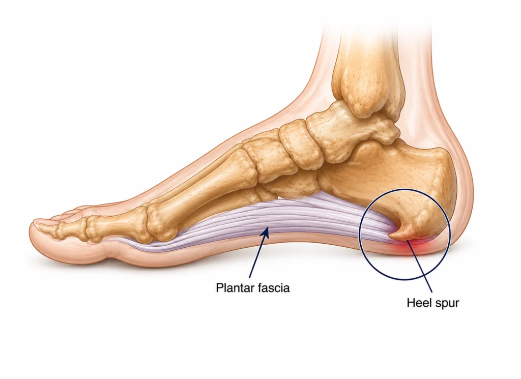
Heel spur or Calcaneal spur are bony growth that can develop at the bottom of heel bone often where the plantar fascia attaches. Posterior heel spur may also develop on the back of heel bone where the achilles tendon attaches.
Heel spur is commonly associated with Plantar fasciitis, but there is no direct link between them. Plantar fasciitis and heel spurs may or may not be present alongside. If present alongside, it usually indicates that the area is under strain for a long time.
At Revamp Healthcare in Dwarka, we help patients across Delhi NCR understand what's causing their heel pain and guide them toward the right treatment, from simple lifestyle changes to custom orthotic insoles designed around their foot's specific structure.
What Causes Heel Spur?
Heel spurs form gradually, as a result of ongoing strain at the point where the plantar fascia and surrounding tissue attach to the heel bone. When this area is repeatedly pulled or stressed over time — much like what happens in long-standing plantar fasciitis — the body responds by laying down small amounts of calcium at that attachment point. Over months, this can build into a small, bony projection.
The tissue at this attachment point isn't designed to handle constant pulling and tension. When it's placed under repeated microscopic strain — the kind that builds up with plantar fasciitis, prolonged standing, or high-impact activity — the fibers in that area gradually break down and are replaced with tougher, less flexible tissue. Over time, this process can trigger calcification, which is how a spur forms. In other words, a heel spur usually reflects months of accumulated strain, rather than a single injury.
Heel spurs are frequently found in people with long-standing plantar fasciitis, since both conditions develop from the same underlying strain on the fascia. However, having one doesn't guarantee the other — some people with significant plantar fasciitis never develop a spur, and some people with a visible spur on an X-ray have no pain at all. Because of this, a heel spur is better understood as a sign of prolonged strain in the area, not something that directly causes pain on its own or needs to be treated separately in most cases.
Common Symptoms
Clinical Examination Includes
A heel spur is usually identified through a combination of your symptom history, a physical examination, and imaging where necessary. Since a spur being present doesn't automatically explain your pain, your specialist's job is to determine what's actually driving your discomfort — whether that's the spur, the surrounding fascia, or another factor entirely.
Treatment & Recovery
Because the pain associated with a heel spur is almost always driven by the surrounding soft tissue — usually plantar fasciitis — rather than the bony growth itself, treatment focuses on calming and correcting that underlying strain, not on removing the spur. The encouraging news is that the vast majority of people improve with non-surgical care alone, though it can take several months of consistent treatment to see full results, and it's worth giving conservative care a fair trial before considering anything more involved.
Reducing the load on the Heel
How Revamp's custom Insoles work for Heel Spur
Custom Insoles is a proven non surgical management of Heel spur and associated symptoms. Custom Insoles Features a deep heel cup which offloads the pressure from the site of spur formation/ inflammation. It also allows reduce strain on the foot by redistributing weight and achieve biomechanically accurate foot alignment.
Night Splints
For people whose pain is worst with the first few steps in the morning, night splints that hold the ankle in a slightly upward-flexed position overnight can meaningfully reduce that first-step pain by preventing the fascia from tightening while you sleep.
When Symptoms doesn't settle with basic treatment
If pain hasn't improved after a couple of months of consistent conservative care, your specialist may recommend a period of more structured immobilisation — such as a walking cast for around six weeks — before transitioning back into orthotics and stretching. This step is uncommon and reserved for cases that haven't responded to the initial approach, not a typical part of first-line treatment.
Reviewed By Dr Mohit Sharma - Clinical Head, Podiatrist, Foot Care Specialist on 22 July 2026